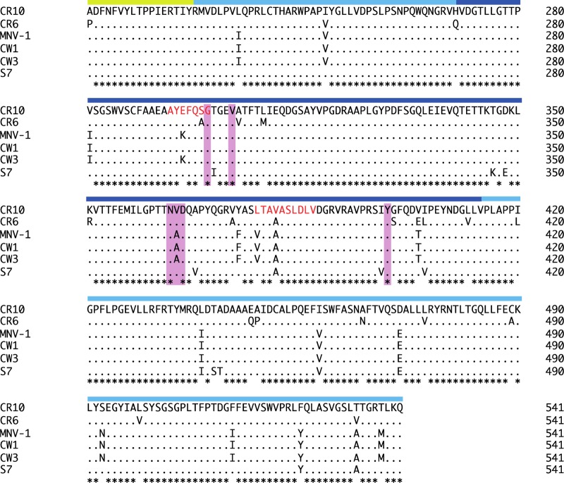
FIG 4.
Sequence alignment of different murine norovirus capsid proteins. The partial S domain (yellow bar), P1 subdomain (light blue bar), and P2 subdomain (dark blue bar) are indicated on the alignment. The CR10 residues (pink highlight) that interacted with the CD300lf through direct hydrogen bonds and hydrophobic interactions were mostly conserved in different murine norovirus strains. The A6.2 antibody recognition site is indicated by red amino acid letters. Asterisks indicate highly conserved residues.

