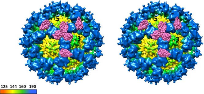
FIG 8.

Cryo-EM map of CW1 murine norovirus virion and CR10 P domain sCD300lf complex structures. Shown is a stereo view of the (cryo-EM) CW1 virion structure superpositioned with the X-ray crystal structure of the CR10 P domain sCD300lf complex. The CR10 P domains are removed for clarity, and two molecules of sCD300lf are shown on each P dimer. The virion was colored according to diameter, showing the S domain (yellow) and the P domain (blue). The sCD300lf slightly clashed at the P dimer intersections, suggesting that multiple sCD300lf molecules might not bind simultaneously at this intersection.
