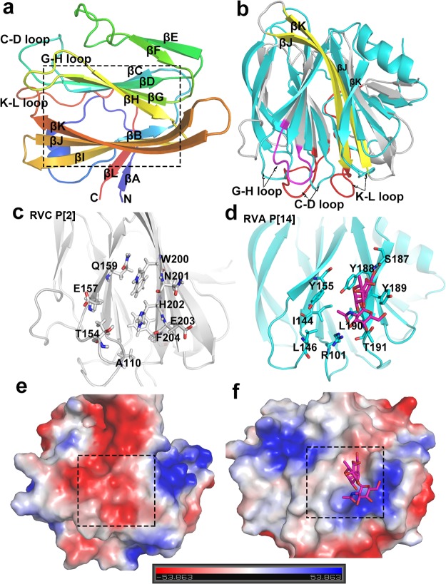
FIG 3.
Crystal structure of the native human RVC VP8* and its comparison with that of the previously reported RVA P[14] VP8* (PDB accession number 4DRV). (a) Cartoon representation of the human RVC VP8* structure showing a galectin-like fold with two twisted β-sheets separated by a cleft (framed in the box). The N and C termini are denoted. (b) Structural alignment of the human RVC VP8* (gray) with the VP8* of human RVA P[14] RVA (cyan). The longer β-strands (βJ, βK), longer loops (C-D loop, K-L loop), and altered G-H loop in RVC VP8* are colored yellow, red, and magenta, respectively (arrows). (c and d) The glycan binding site in the human RVA P[14] VP8* (cyan) (d) and the corresponding site in the human RVC VP8* (gray) (c) (cartoon representation) with indications of the related amino acids (stick representation). (e and f) Comparison of the electrostatic surface potentials of the glycan binding sites between the human RVC VP8* (e) and the human RVA P[14] VP8* (f). Both VP8*s are shown in surface representation. The blue and red indicate the positive and negative electrostatic surface potentials, respectively. The glycan binding sites are framed by dashed-line boxes.

