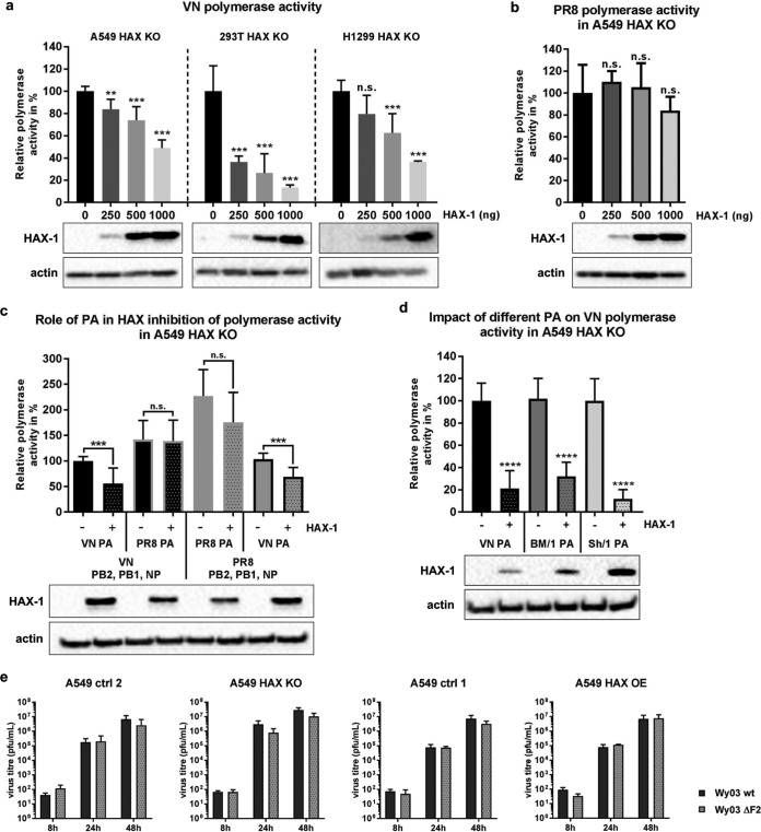
FIG 6.
HAX-1 inhibits viral polymerase activity in a PA-dependent manner. (a to d) Mini-replicon assays were performed in A549, H1299, and 293T cells lacking HAX-1 expression. Cells were transfected with a plasmid carrying a firefly luciferase-based minigenome and one encoding Renilla luciferase under the control of a constitutive promoter, as well as the following plasmids: 10 ng of pCAGGS.VN-PB2, 10 ng of pCAGGS.VN-PB1, 10 ng of pCAGGS.VN-PA, 20 ng of pCAGGS.VN-NP, and different amounts of pCAGGS.HAX-1 (a); 10 ng of pCAGGS.PR8-PB2, 10 ng of pCAGGS.PR8-PB1, 10 ng of pCAGGS.PR8-PA, 20 ng of pCAGGS.PR8-NP, and different amounts of pCAGGS.HAX-1 (b); either 10 ng of pCAGGS.VN-PB2, 10 ng of pCAGGS.VN-PB1, 20 ng of pCAGGS.VN-NP, or 10 ng of pCAGGS.PR8-PB2, 10 ng of pCAGGS.PR8-PB1, and 20 ng of pCAGGS.PR8-NP with the 10-ng PA segment of either pCAGGS.VN-PA or pCAGGS.PR8-PA and with or without 500 ng of pCAGGS.HAX-1 (c); and 10 ng of pCAGGS.VN-PB2, 10 ng of pCAGGS.VN-PB1, and 20 ng of pCAGGS.VN-NP with the 10-ng PA segment of pCAGGS.VN-PA or pCAGGS.BM/1-PA or pCAGGS.Sh/1-PA (d). Cells were lysed 24 h posttransfection, and lysates were subjected to luciferase assay. Values represent means from triplicate experiments, with SDs. One-way ANOVA was used. **, P < 0.01; ***, P < 0.001; ****, P < 0.0001. Cell lysates were also used for WB to visualize HAX-1 expression and actin. (e) Different A549 cell lines were infected at an MOI of 0.01 with either Wy/03 wt or Wy/03 ΔF2 virus with TPCK-treated trypsin at 0.1 μg/ml. Supernatants were collected at the indicated time point postinfection, and virus titer was assessed by plaque assay on MDCK cells. Bars represent means (n = 3), with SDs.

