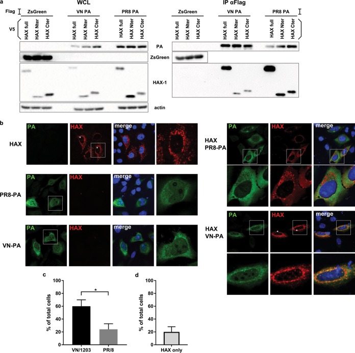
FIG 7.
HAX-1 interacts with the PA subunit of the polymerase. (a) 293T cells were transfected with each indicated protein cloned into the mammalian expression plasmid pCAGGS.V5-HAX-1 and pCAGGS.Flag-VN-PA. Cell lysates were collected 48 h later and processed through immunoprecipitation using a monoclonal antibody against Flag-tagged proteins. Samples were then analyzed by Western blotting. (b) HeLa cells were transfected with V5-HAX-1 and Flag-PA proteins from either VN/1203 or PR/8. Cells were fixed at 48 h and processed for detection of HAX-1 (red) and PA (green) by immunofluorescence and stained with DAPI. An asterisk indicates cells where HAX-1 is present in the cytoplasm. (c and d) Graphs represent the percentages of cells where HAX-1 localizes to the nucleus. Bars represent the means, with SEMs (n = 25). (c) In samples transfected with both HAX-1 and PA, we took into account cells that were doubly positive. *, P < 0.05. (d) In samples transfected with HAX-1 only, we took into account every HAX-1-positive cells.

