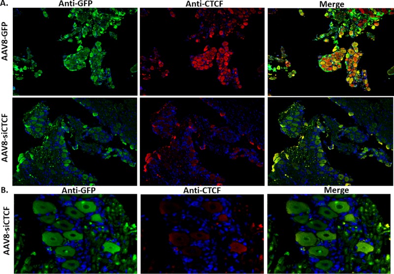
FIG 3.
Analysis of rabbit TG neurons for AAV transduction and CTCF expression. Rabbits were treated with AAV vectors on the eye. Two weeks later, rabbits were sacrificed, and the TG were removed and sectioned for immunofluorescence. The sections were incubated with mouse anti-GFP (primary) and Alexa Fluor 488-conjugated (secondary) antibodies for the visualization of the GFP-expressing AAV8 (AAV8-GFP) and mouse anti-CTCF (primary) and an Alexa Fluor 548-conjugated (secondary) antibodies for the visualization of CTCF. DAPI staining in blue represents nuclei of satellite cells present in the sections. Panel A images are shown at ×10 magnification. Panel B images show only AAV-siCTCF-treated animals at a ×40 magnification.

