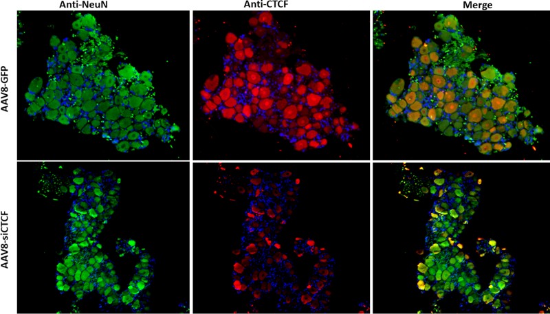
FIG 5.
Immunofluorescence was performed to show neuronal colocalization of the AAV8-GFP, NeuN, and CTCF. The sections were incubated with mouse anti-NeuN (primary) and an Alexa Fluor 488-conjugated (secondary) antibodies for the visualization of the NeuN and mouse anti-CTCF (primary) and Alexa Fluor 548-conjugated (secondary) antibodies for the visualization of CTCF. DAPI staining in blue represents nuclei of satellite cells present in the sections. Images are shown at ×10 magnification.

