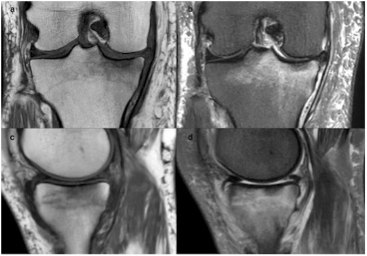
Fig. 3.
Comparison of conventional MRI on coronal T1 and STIR (a, b) vs. synthetic STIR on sagittal T1 and STIR (c, d) for a 45-year-old patient consulting for internal knee pain. For this particular example, the conventional STIR was acquired also in the sagittal orientation (usually in coronal). The edema is clearly visible on the tibial endplate on synthetic imaging.

