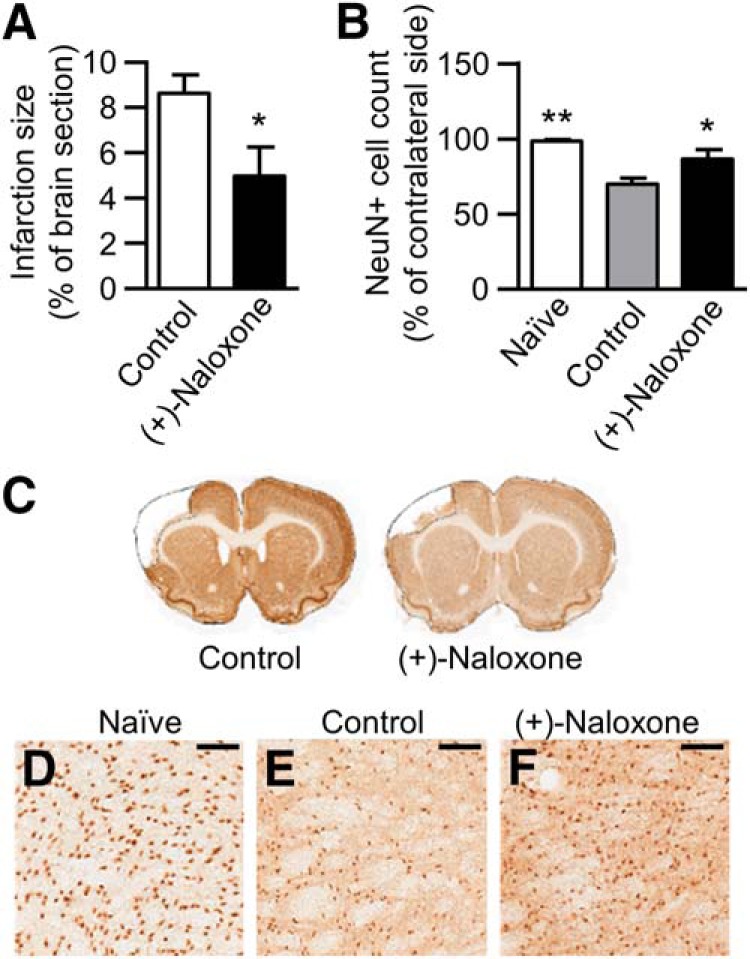
Figure 6.

Post-stroke intranasal (+)-naloxone decreases infarction area and neuronal loss in the thalamus. A, Average infarction size calculated from NeuN-negative area at day 14 post-stroke. *, p < 0.05, Student’s t test. B, Average number of neurons (NeuN+ cells) in the ipsilateral thalamus at day 14 post-stroke expressed as a percentage of the contralateral thalamus. *, p < 0.05 and **, p < 0.01 indicate pairwise comparison with the control group with Mann–Whitney U test following Kruskal–Wallis test. C, Representative photomicrographs of anti-NeuN immunostained brain sections, with infarction area delineated. D–F, Representative photomicrographs of anti-NeuN immunostaining of ipsilateral thalamus in naive (D), control (E), and (+)-naloxone–treated (F) rats. Scale bar is 150 µm. Naive, no-stroke rats (n = 6); control, stroke rats with vehicle or no treatment (n = 18); (+)-naloxone, 0.32–0.8 mg/kg (n = 10). The data represent mean ± SEM.
