Figure 7.
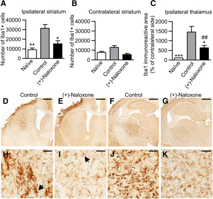
Post-stroke intranasal (+)-naloxone decreases microglia/macrophage activation in the striatum and thalamus. A, B, Microglia/macrophages (Iba1+ cells) were counted with unbiased stereology in the ipsilateral (A) and contralateral (B) striatum. *, p < 0.05 and **, p < 0.01 indicate pairwise comparison with the control group with Bonferroni’s post hoc test following one-way ANOVA. C, The area of Iba1+ cells in the ipsilateral thalamus expressed as a percentage of the contralateral thalamus. *, p < 0.05 and ***, p < 0.001 indicate pairwise comparison with the control group; ##, p < 0.01 indicates comparison with the naive group with Mann–Whitney U test following Kruskal–Wallis test. D–K, Representative photomicrographs of anti-Iba1 immunostaining of ipsilateral striatum (D, E) and thalamus (F, G) in control (D, F) and (+)-naloxone–treated (E, G) rats; H–K show high magnification. Black arrow shows a typical Iba1+ cell. Scale bars are 1000 µm (D–G) and 50 µm (H–K). Naive, no-stroke rats (n = 6); control, stroke rats with vehicle or no treatment (n = 18); (+)-naloxone, 0.32–0.8 mg/kg (n = 10). The data represent mean ± SEM.

