FIG 2.
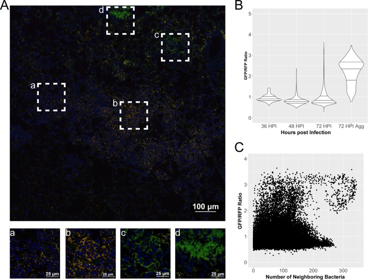
Expression of crp is increased in large aggregates or biofilm-like structures in the lungs. Cross sections of lungs from mice infected with strain CO92 carrying the Pcrp-GFP reporter (green) and pGEN-RFP plasmid (red) were stained with DAPI (blue) and imaged by confocal microscopy. (A) Large image generated with the tile feature in NIS Elements acquisition software to cover Y. pestis-containing lesions within the lung space. Magnified images of boxed areas (a to d) highlight heterogeneity in expression patterns of crp within a single lesion. (B) Violin plots displaying the relative expression of crp as a ratio of GFP to RFP, quantified in individual cells combined from the lung lesions of at least three mice at each time point, across two independent infection experiments at 36, 48, and 72 hpi. Horizontal lines within the violins represent the 25th percentile, median, and 75th percentile. Aggregates were also imaged and analyzed separately. (C) Expression of crp in individual cells as a function of neighboring bacterial cell density from all images and time points postinfection.

