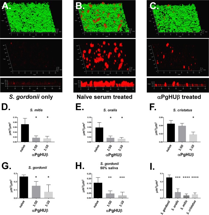
FIG 3.
Preincubation of P. gingivalis with anti-PgHUβ prevents P. gingivalis from entering into streptococcal biofilm. Biofilms were grown for 24 h before addition of P. gingivalis. P. gingivalis was incubated with a 1:50 or 1:10 dilution of anti-PgHUβ antisera for 1 h before addition. Representative biofilm images show results for S. gordonii alone (A) as well as naive serum (B) and a 1:50 dilution of anti-PgHUβ-treated P. gingivalis (C). All cells (green) and P. gingivalis (red) are shown in the top image, while P. gingivalis-only labeling is shown in in the middle image. The bottom image is a side view of the P. gingivalis-only-labeled biofilm. A no-P. gingivalis control biofilm (A) shows little to no red signal, while that treated with naive serum shows significant amounts of P. gingivalis (B). Treatment with a 1:50 dilution of anti-PgHUβ results in a significant decrease in P. gingivalis detected within the biofilm (C). Graphical representations of decrease in P. gingivalis entering into S. mitis (D), S. oralis (E), S. cristatus (F), or S. gordonii (G) indicate dose-dependent decreases in detection of P. gingivalis. Experiments with S. gordonii were repeated with 50% pooled human saliva (H). Total naive serum-treated P. gingivalis entering streptococcal biofilms is shown in panel I, which shows that significantly more P. gingivalis cells enter an S. gordonii biofilm than the other oral streptococci tested. Dual-species biofilms were grown for 16 h in THBHK. Cells were stained with carboxyfluorescein succinimidyl ester (CFSE). P. gingivalis was immunofluorescently labeled with a polyclonal antifimbrial primary antisera and a secondary goat anti-rabbit Alexa Fluor 647-conjugated antibody and visualized using confocal scanning laser microscopy. *, P ≤ 0.05; **, P ≤ 0.01; ***, P ≤ 0.001.

