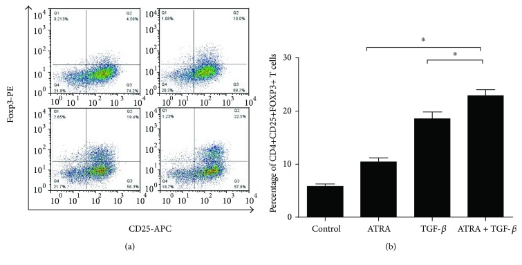
Figure 1.
The percentage and number of Tregs in SSc CD4+ T cells. (a) SSc CD4+ T cells were treated with 10 nm ATRA and/or 10 ng/ml TGF-β in the presence of anti-CD3/CD28 beads and 200 U IL-2 for 4 days. CD4+CD25+FOXP3+ T cells were analyzed by flow cytometry. (b) The bar graph shows the percentage of CD4+CD25+FOXP3+ Tregs in samples of SSc in different groups. (b) The proportion of CD4+CD25+FOXP3+ Tregs was significantly increased in SSc CD4+ T cells treated with ATRA and/or TGF-β compared with the blank control group (all, p < 0.0.05). The ATRA and TGF-β combined stimulus group showed a significantly increased percentage of Tregs compared with the ATRA or TGF-β alone group (∗p < 0.05). Data are presented as the mean ± SD and are representative of at least 3 independent experiments.

