
Figure 4. Paramyxoviruses produced in SPCA1 KO cells are less infectious.
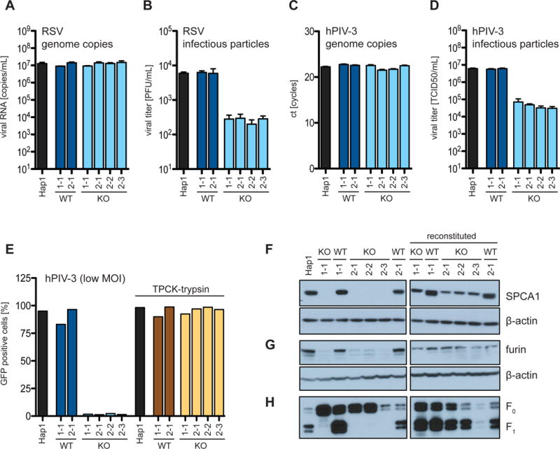
(A and B) Hap1 cells infected with RSV-GFP at a MOI of 1 and (C and D) with hPIV-3-GFP at a MOI of 4. The supernatants were harvested at 24 hpi and viral genome copies determined by (A and C) qPCR and infectious particles by (B) plaque assay and (D) TCID50. (E) Hap1 cells were infected with hPIV-3-GFP at a MOI of 0.01. Cells were cultured in low serum conditions (0.1% FBS) and supplemented as indicated with TPCK-trypsin (1 μg/ml). (A-D) Data represent the mean and SD of 3 independent experiments. Cells are colored differently to indicate: parental WT = black; CRISPR WT clones = dark blue and brown; and CRISPR KO clones = light blue and yellow. (F and G) Western blot analysis of Hap1 cells, CRISPR-generated Hap1 clones and SPCA1-reconstituted clones for (F) SPCA1 and (G) furin. (H) Infection of Hap1 cells, CRISPR-generated Hap1 clones and SPCA1-reconstituted clones with hPIV-3-GFP at a MOI of 2. Viral particles released into supernatants were harvested at 24 hpi and probed by western blot for the viral fusion protein F in its uncleaved F0 and cleaved F1 conformation.