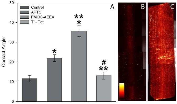
Figure 2. Evidence of chemical modification of surfaces.
(A) Contact Angle Analysis: Using the sessile drop technique, surface contact angles of a 10μL droplet of dH2O was measured on control Ti-OH discs (following autoclaving), silanized Ti discs (APTS), silanized Ti discs with attached Fmoc-AEEA linkers (Fmoc-AEEA) and the completed covalently tethered tetracycline (Ti-TET). Data are presented as means and standard error. A one-way analysis of variance test was used and a statistically significant difference (p < 0.05) was found between control and both APTS and Fmoc-AEEA (*), between APTS and both Fmoc-AEEA and Ti-TET (**), as well as between Fmoc-AEEA and Ti-TET (#). Immunohistological Analysis: Ti control rods (B) and those tethered with tetracycline (C) were each examined using an antibody specific against the 4-dimethylamino group of tetracycline. Note the minimal fluorescence on the control rods in comparison to the extensive level of fluorescence on Ti-TET rods. When a similar assay was performed using an antibody specific to the 2-(amino-hydroxy-methylidene) group of tetracycline (the theoretical binding site), minimal fluorescence was observed on both surfaces. Fluorescence is presented as an intensity map (see legend in B).

