Fig. 4.

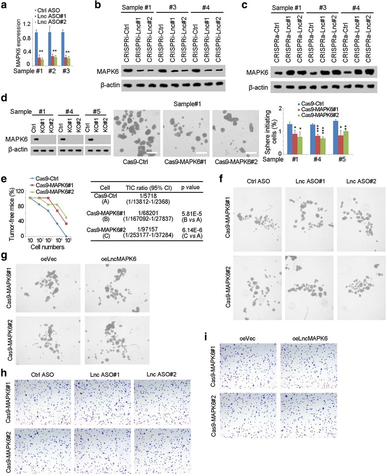
LncMAPK6 drove MAPK6 expression. (a) MAPK6 expression in lncMAPK6 depleted cells was detected using three primary samples. (b, c) LncMAPK6 depleted (b) and overexpressed (c) cells were used for MAPK6 examined with immunobloting. CRISPRi-lnc, CRISPRi-lncMAPK6; CRISPRa-lnc, CRISPRa- lncMAPK6. β-actin was a loading control. (d) MAPK6 knockout cells were established and sphere formation assay was performed. Typical images and indicated ratios were shown in middle and right panels, respectively. (e) 10, 1 × 10², 1 × 10³, 1 × 10⁴ and 1 × 10⁵ MAPK6 knockout cells were used for tumor formation. (f, g) LncMAPK6 was silenced (f) or overexpressed (g) in MAPK6 knockout cells and lncMAPK6 didn’t participate in liver TIC self-renewal upon MAPK6 was deleted. (h, i) MAPK6 knockout cells were used for lncMAPK6 knockdown (h) or overexpression (i), followed by transwell assay
