Fig. 2.

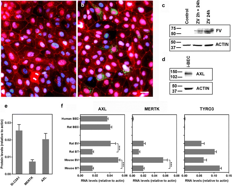
ZIKV infection and AXL expression in BECs. a, b Immunofluorescence staining was conducted 24 h post ZIKV infection (MOI = 4) of i-BECs using anti-flavivirus group antibody (FV; green). ZIKV was detected in infected i-BECs (b) but not in uninfected controls (a). Tight junction marker ZO-1 was used to label cell–cell contacts (red); nuclei were stained using Hoechst dye (blue). Scale bar = 10 µm. c The presence of ZIKV in i-BECs was also determined by Western blotting at 2 or 24 h after exposure to the virus. d Representative Western blot demonstrating putative ZIKV receptor AXL expression in i-BECs using an anti-AXL antibody. β-ACTIN was used as an internal loading control. e Expression levels of SLC2A1 (GLUT-1), MERTK and AXL in human brain endothelial cell line hCMEC/D3 determined by proteomics (LC–MS). Shown are ratio of intensity-based absolute quantification values (mean ± SD from 3 independent cell preparations) calculated for each protein and β-ACTIN, as described previously [41]. f The expression of AXL, MERTK and TYRO3 in primary human and rat brain endothelial cells (BECs) or in isolated rat or mouse brain vessels (BV) or whole brain tissues (BT), determined by NGS. Shown are abundances (mean ± SD) of RNA transcripts relative to β-ACTIN from at least three cell/tissue preparations (***p < 0.001, One-Way ANOVA, Tukey’s post hoc test)
