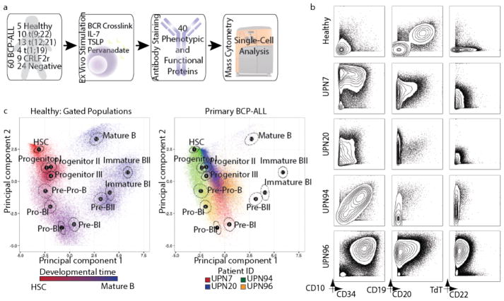
Figure 1. Mass cytometry analysis of BCP-ALL reveals phenotypic heterogeneity of leukemic cells.
(a) Summary of primary BCP-ALL sample processing for mass cytometry analysis (see Supplementary Tables 1–3 for patient information, antibody panel, and perturbation conditions, respectively). 60 primary BCP-ALL samples and 5 healthy control bone marrow aspirates were included. Prognostic cytogenetic translocations identified at diagnosis, as well as relevant ex vivo perturbations used to uncover cell state, are indicated. ‘Negative’ patients were negative for any of the prognostic cytogenetic translocations analyzed. (b) Mass cytometry analysis of commonly used diagnostic antigens expressed by lineage-negative bone marrow cells (see Supplementary Fig. 1a for gating) from 4 representative BCP-ALL patients and 1 healthy donor. (c) Left panel: 5,000 cells from 12 manually gated stages of B-cell development in healthy bone marrow demonstrate phenotypic progression during normal B lymphopoiesis (1,000 cells sampled from each of n = 5 donors). The first 2 principal components were constructed using 11 markers defining B-cell developmental populations (see Supplementary Figs. 1b–d for gating, marker weights, and variance captured by each principal component). The developmental time color scale was defined by setting hematopoietic stem cells as red and mature B cells as blue. Intermediate populations were placed on this red-to-blue color gradient at equal intervals. For each stage, a black dot indicates the population centroid, and the surrounding circle indicates standard error based on 5 healthy donors. Right panel: Data from 4 patients in (b) shown projected onto healthy B-cell progression. Each sample uniquely occupies the PCA space, while overlapping with multiple healthy populations and other patient samples. BCR, B-cell receptor; TSLP, thymic stromal lymphopoietin; PCA, principle component analysis.

