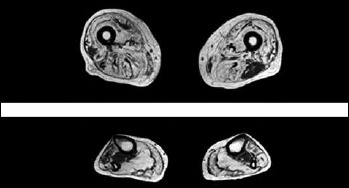
Figure 3.

Muscle MRI of thigh and lower legs showing extensive T1w hyperintensity in most muscle groups suggesting severe fatty and/or fibrous degeneration. At thigh level there is an asymmetric relative sparing of the left biceps femoris and to a lesser extent of the left gracilis with a severe involvement of the other muscles and a characteristic patchy appearance particularly in vastus lateralis. At lower leg level there is also a severe involvement of both anterior and posterior compartment with a patchy appearance of anterior tibialis and peroneal muscles and a relative sparing of tibialis posterior muscle.
