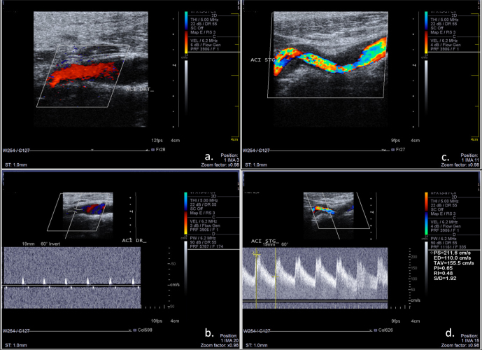
Figure 2.
Ultrasound examination of the carotid arteries. a: color mode examination of the right ICA, longitudinal section, without stenotic lesions; b: real-time triplex display (Color Doppler+ Pulsed Spectral Doppler) of the right ICA revealing high-resistance flow signal, with low and short systolic flow and completely absent diastolic flow, indicative of near occlusion or occlusion of the distal segment of ICA; c: color-mode examination of the left ICA revealing irregular stenosis caused by the hypoechoic mural hematoma; d: triplex mod examination of the left ICA, revealing high flow velocities suggestive of significant stenosis.

