Figure 4.
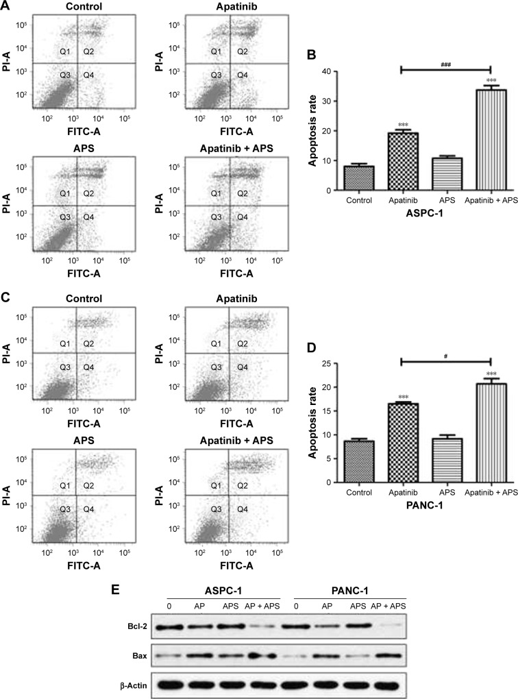
APS increased apoptosis induced by apatinib in pancreatic cancer cells.
Notes: ASPC-1 (A and B) and PANC-1 (C and D) exposed to control, 40 μM apatinib, 200 μg/mL APS, and 40 μM apatinib + 200 μg/mL APS after 24 hours followed by Annexin V-FITC and PI staining, and apoptosis percentage was detected by flow cytometry. (E) Proapoptotic (Bax) and antiapoptotic (Bcl-2) proteins expression treated with 0 (control), AP (40 μM apatinib), APS (200 μg/mL) and AP + APS (40 μM apatinib + 200 μg/mL APS) after 24 hours were determined by Western blotting. β-Actin was used as the internal control. ***P<0.001 versus control; #P<0.05, ###P<0.001 versus apatinib.
Abbreviations: AP, apatinib; APS, Astragalus polysaccharide; FITC, fluorescein isothiocyanate; PI, propidium iodide.

