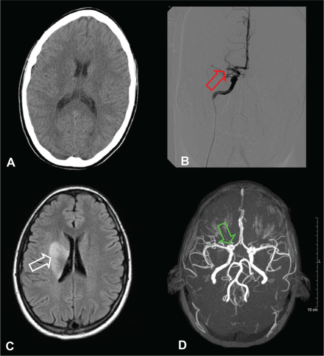
Figure 1.
Cranial CT at 60 minutes after the stroke onset without any acute intracranial ischemic lesion or hemorrhage (image A); Angiography at 120 minutes after stroke onset showed complete occlusion of right middle cerebral artery (image B, red arrow); Cranial MRI at seven days after stroke onset and intra-arterial thrombolysis showing ischemic lesion of right sided basal ganglia (image C, white arrow); Cranial MR angiogram with intact Willis circle and complete recanalization of the right middle cerebral artery (image D, green arrow).

