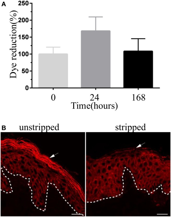
Figure 1.

Validation of human skin explant model. (A) The viability of human skin explant in culture. Each bar represents the reduction of PrestoBlue dye staining (a cell viability testing reagent) by skin samples at different time points in culture compared with freshly received skin tissue (time 0). Results are pooled from three independent experiments. (B) Immunofluorescence images showing skin cryosections of unstripped and stripped human skin tissue stained with anti-pan-cytokeratin antibody. Stripped skin lacks stratum corneum (arrow). Scale bar: 20 µm.
