Figure 6.
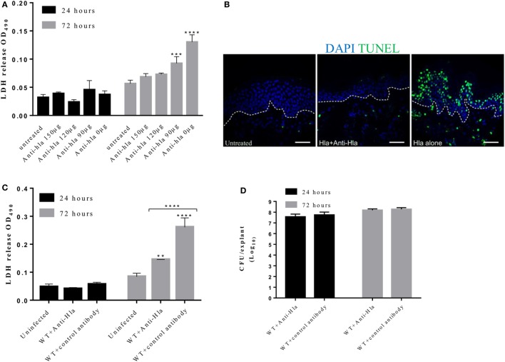
Anti-Hla antibodies mitigate tissue cytolysis but not bacteria survival during infection. Human skin explants were treated with 5 µg of Hla in combination with increasing concentrations of rabbit anti-Hla antibodies. For infection experiments, skin explants were infected with 5 × 10⁵ colony-forming unit (CFU) of Staphylococcus aureus USA300 wild type in combination with 150 µg of purified rabbit anti-Hla antibodies or unspecific antibodies. (A) Inhibition of purified Hla activity by anti-Hla antibodies in a concentration-dependent manner. Each bar represents the mean ± SD (N = 3). (B) Immunofluorescence images of skin sections showing TUNEL-positive cells in the skin treated with Hla in combination with 150 µg of anti-Hla antibodies. White dotted lines represent the boundary between the epidermis and dermis. Scale bar is 50 µm. (C) Lactate dehydrogenase (LDH) content in the medium of infected skin. Each bar represents the mean ± SD (N = 3). (D) Bacteria CFU recovered from the skin 24 and 72 h post-infection. Each bar represents the mean ± SD (N = 3). Statistically significant differences were determined by two-way ANOVA, with Tukey’s multiple comparison tests, **p ≤ 0.05, ***p ≤ 0.0002, and ****p < 0.0001.

