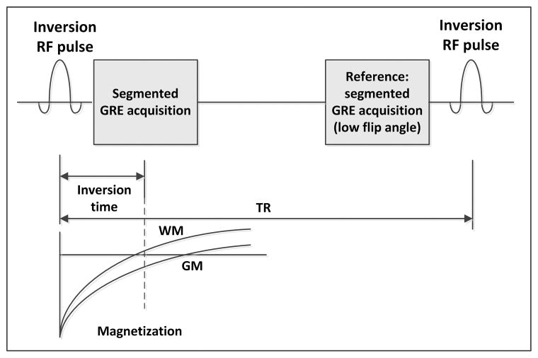
Figure 1.
Pulse sequence diagram of the 2D PSIR acquisition: a non-selective inversion radio frequency (RF) pulse is applied and after an inversion time (TI) a segment of a 2D gradient echo image (GRE) is acquired. The same segment, used as reference for the phase sensitive reconstruction, is reacquired with a low flip angle just before the application of a subsequent inversion RF pulse and the acquisition of the following segment. The time between RF inversion pulses is called repetition time (TR). The magnetization in most of the tissues can be considered almost fully recovered when the reference image is acquired.

