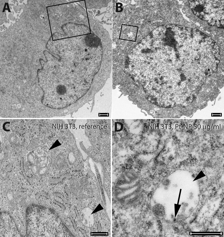
Fig 10. Ultrastructure of NIH 3T3 cells following exposure to Pt-NP.
NIH 3T3 cells were cultivated either without any additional Pt particles as reference (A, insert in C) or in culture medium containing 50 μg/ml (B, insert in D) of the Pt-NP. No cytotoxic effect of Pt-NP could be found. Fibroblasts were highly active in endocytosis (rectangles in A and B). In the multi-vesicular bodies (arrowheads in C and D) accumulation of adsorbed material such as Pt-NP (arrow in D) was detected. Size of bars: 1 μm.

