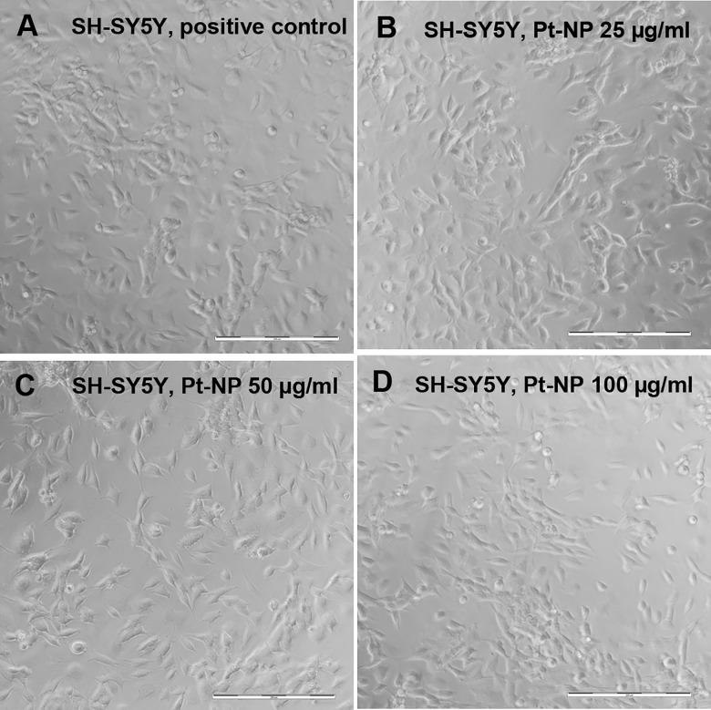
Fig 11. Microscopic characterization of the morphology of SH-SY5Y cells following exposure to Pt-NP.
SH-SY5Y cells were cultivated either without any additional Pt particles as reference (A) or in culture medium containing 25 μg/ml (B), 50 μg/ml (C) and 100 μg/ml (D) of the Pt-NP. Morphology and adhesion behavior of the SH-SY5Y cells did not change throughout the cultivation assays at varying Pt-NP concentrations. Size of bars: 200 μm.

