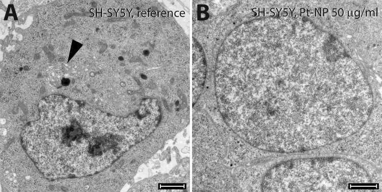
Fig 12. Ultrastructural morphology of SH-SY5Y cells following exposure to Pt-NP.
SH-SY5Y cells were cultivated either without any additional Pt particles as reference (A) or in culture medium containing 50 μg/ml (B) of the Pt-NP. The biosynthesis of synaptic granules as seen in the control cells around the Golgi apparatus (arrowhead in A) is typical for those cells; no multi-vesicular bodies of the endocytosis were detected. Neuroblastoma cells in contact with Pt-NP were active only in the secretory pathway. No Pt-NP were evident inside the cell. Size of bars: 200 μm.

