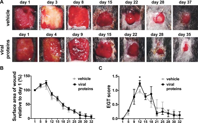
Fig 1. Healing of limb wounds of horses following treatment with viral proteins.
(A) Representative photos of healing limb wounds following a single topical administration of vehicle (HyStem hydrogel) or viral proteins in vehicle (VEGF-E and ovIL-10) prior to bandaging on day 1 and a subcutaneous injection of vehicle (saline) or viral proteins in vehicle at day 8. (B) Surface area and (C) exuberant granulation tissue (EGT) formation of healing wounds at the days indicated. Wound surface area is calculated relative to the original wound area. EGT formation was scored on 50% on protuberance (0 none– 2 marked), 25% colour (0 pink– 1 yellow-red) and 25% on quality (0 smooth—1 rough). Values represent mean ± SEM, n = 4. *p ≤ 0.05 between means of viral protein treated and vehicle control wounds at each time point, as determined using a linear mixed model with a priori contrasts followed by a Benjamini-Hochberg sequential adjustment.

