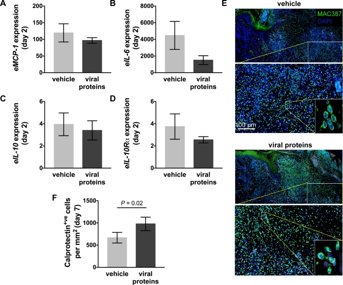
Fig 3. Inflammation in limb wounds of horses following treatment with viral proteins.
Quantitative PCR was used to measure the expression of (A) equine (e)MCP-1, (B) eIL-6, (C) eIL-10, and (D) eIL-10Rα in wound margin samples taken 2 days after wounding. Expression of mRNA is relative to that of GAPDH and to levels measured in unwounded skin (day 0). (E) Representative photos of skin sections taken 7 days post-wounding stained with DAPI (blue) and an antibody against the inflammatory cell marker calprotectin (MAC387: green). Enlarged images show nucleated inflammatory cells. (F) Number of calprotectin+ve cells in the granulation tissue and surrounding skin of wounds following administration of vehicle or viral proteins. Values represent mean ± SEM, n = 4. P values indicated were determined using a two-tailed ratio paired t-test.

