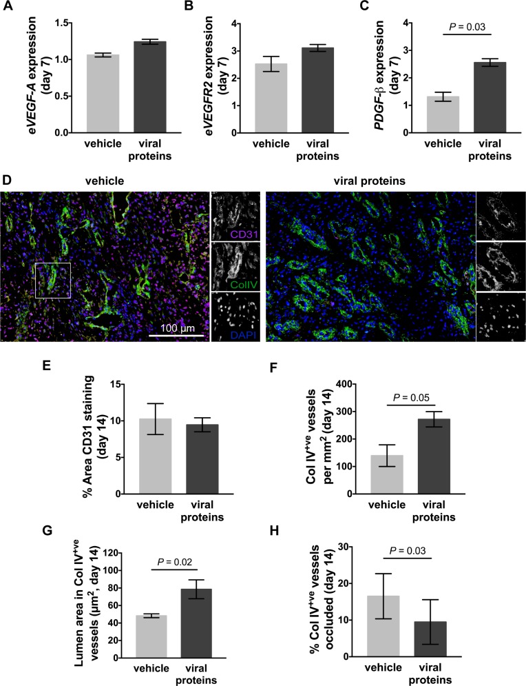
Fig 4. Vascularisation of limb wounds of horses following treatment with viral proteins.
Quantitative PCR was used to measure the expression of (A) eVEGF-A, (B) eVEGFR2, and (C) ePDGF-β in wound margin samples taken 7 days after wounding. Expression of mRNA is relative to that of GAPDH and to levels measured in unwounded skin (day 0). (D) Representative photos of skin sections taken 14 days post-wounding, stained with DAPI (blue) and antibodies against blood vessel endothelial cells (CD31: pink) and basal lamina (collagen IV: green). Enlargements show single colour images of representative blood vessels. (E) Area of CD31 staining, (F) number of collagen IV+ve blood vessels, (G) lumen area in collagen IV+ve blood vessels and (H) percentage of blood vessels occluded (lumen area less than 7 μm2) in the granulation tissue of wounds following administration of vehicle or viral proteins. Values represent mean ± SEM, n = 4. P values indicated were determined using a two-tailed ratio paired t-test.

