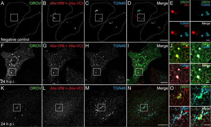
Fig 7. Alix is recruited to TGN during OROV assembly.
Mock infected (A-D) or OROV infected (F-I) HeLa cells were transfected with VCt-Alix and VNt-Alix plasmids (red). Cells were immunostained to OROV proteins (green) and TGN46 (cyan) and analyzed by confocal microscopy. (K-N) 3D-SIM images of HeLa cells processed as (F-I). The image represents a projection of Z stacks (125 μm each) of cells after deconvolution. Bars = 10 μm. (E, J and O) Insets representing the boxed areas of A-D, F-I and K-N, respectively. Bars = 2 μm.

