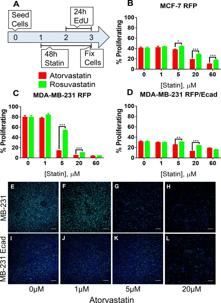
Fig 2. Atorvastatin decreases proliferation of breast cancer cells more potently than rosuvastatin.
(A) Experimental schematic for assessing the proliferation of breast cancer cells under treatment with atorvastatin or rosuvastatin for 48 hours. (B) MCF-7 RFP, (C) MDA-MB-231 RFP, and (D) MDA-MB-231 RFP/Ecad were cultured with atorvastatin or rosuvastatin for 48 hours; during the final 24 hours the media included 10uM EdU. Cells were fixed, EdU was detected, and cells were counterstained with DAPI to label all nuclei. Cellular proliferation was quantified by determining the percentage of EdU positive cells (green, all nuclei are blue—DAPI). (E-H) MDA-MB-231 RFP and (I-L) MDA-MB-231 RFP/Ecad cells treated with (E,I) 0μM, (F,J) 1μM, (G,K) 5μM, or (H,L) 20μM atorvastatin demonstrate both E-cadherin mediated growth suppression and atorvastatin resistance. All data are representative of at least three independent experiments. * P < 0.05, ** P < 0.01, *** P < 0.001.

