. 2018 May 15;7:e33953. doi: 10.7554/eLife.33953

Figure 5. Sequence similarity network (SSN) analysis reveals that the gut bacterial enzyme Cgr2 is a highly distinct member of a large enzyme family that is widespread in gut microbes.

The SSN was constructed using the top 5000 most similar proteins to Cgr2 from the UniprotKB database. Nodes represent proteins with 100% sequence identity. (A) SSN displayed with an e-value threshold of 10−50. The seven previously characterized enzymes (PDB ID: 1D4D, 1E39; UniProtKB ID: Q07WU7, Q9Z4P0, 8CVD0, P71864, Q7D5C1) and Cgr2 are colored according to biochemical function. (B) SSN displayed with an e-value threshold of 10−130. All nodes that co-clustered with characterized enzymes are shown in the same color, denoting putative isofunctional activity. With the exception of Cgr2, if a node comes from a gut bacterium, it is colored red rather than the color of the corresponding cluster.

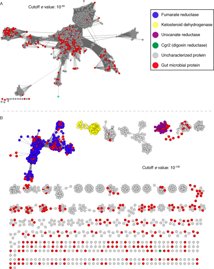
Figure 5—source data 1. Characterized enzymes within the Cgr2 sequence similarity network.
DOI: 10.7554/eLife.33953.031

Figure 5.

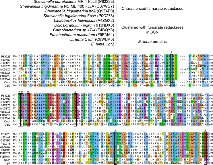
Figure 5—figure supplement 1. Multiple sequence alignment of fumarate reductases.

Figure 5—figure supplement 1.

UniProtKB ID numbers are shown in parentheses. Active site residues (marked with an asterisk) were conserved in characterized fumarate reductases and clustered proteins from the sequence similarity network. These residues were not conserved in Cgr2 and another predicted fumarate reductase (Cac4) associated with the cgr gene cluster.
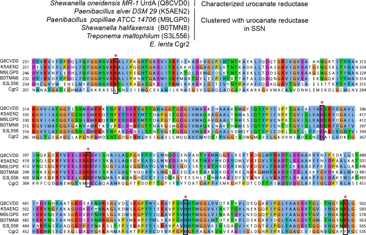
Figure 5—figure supplement 2. Multiple sequence alignment of urocanate reductases.

Figure 5—figure supplement 2.

UniProtKB ID numbers are shown in parentheses. Active site residues (marked with an asterisk) were conserved in characterized urocanate reductases and clustered proteins from the sequence similarity network and were not conserved in Cgr2.
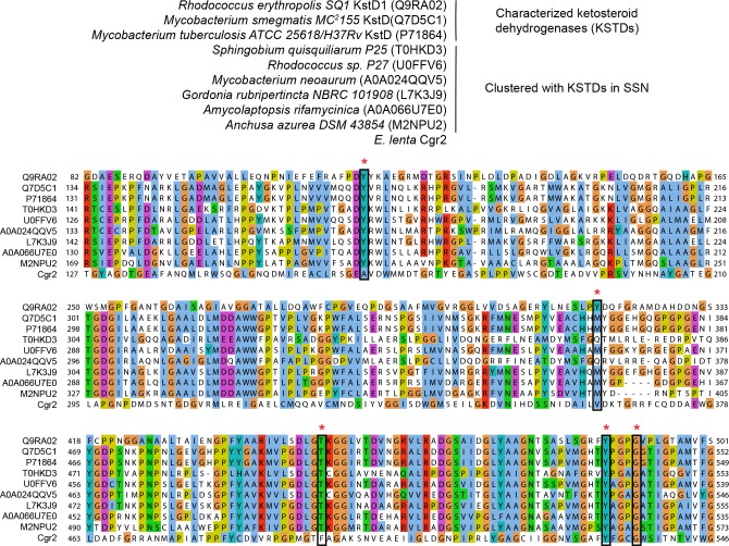
Figure 5—figure supplement 3. Multiple sequence alignment of ketosteroid dehydrogenases.

Figure 5—figure supplement 3.

UniProtKB ID numbers are shown in parentheses. Active site residues (marked with an asterisk) were conserved in characterized ketosteroid dehydrogenases and clustered proteins from the sequence similarity network. Two residues involved in substrate binding and activation were conserved in Cgr2 (Y532, G536).
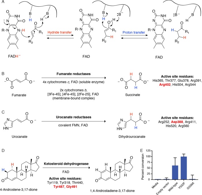
Figure 5—figure supplement 4. Cgr2 is a distinct flavoprotein reductase.

Figure 5—figure supplement 4.

(A) General mechanism of catalysis by Cgr2 homologs. Cgr2 appeared to lack most of the conserved active site residues found in the most similar related enzymes, including (B) 6/7 residues utilized by fumarate reductases, (C) 4/5 residues utilized by urocanate reductases, and (D) 3/5 residues utilized by ketosteroid dehydrogenases. Active site residues are shown with numbering based on S. putrefaciens fumarate reductase, S. oneidensis MR-1 urocanate reductase, and R. erythropolis SQ1 ketosteroid dehydrogenase. Residues shown in red were conserved in Cgr2. (E) Two residues involved in substrate binding and activation in ketosteroid dehydrogenases were conserved in Cgr2 (Y532, G536). Whole cell assays in R. erythropolis overexpressing putative active site mutants in Cgr2 showed that Y532 was not essential for Cgr2 activity towards digoxin. Data represents mean ± SEM (n = 3 biological replicates).