. 2018 Apr 26;7:e34292. doi: 10.7554/eLife.34292

Figure 2. Maximum-likelihood phylogeny of RquA proteins constructed from an alignment of homologs from 166 organisms and 197 aligned sites.

Eukaryotic proteins are coloured based on their phylogenetic affiliations: Obazoa (purple), Stramenopiles-Alveolata (orange), Excavata (green), Amoebozoa (magenta) and Rhizaria (blue). Hexagons represent taxa where RQ has been detected experimentally. Proteobacterial designations (α,β,γ) are indicated in the grey squares. Genetic linkage of rquA and genes related to respiratory function (complex I-IV, cytochrome c metabolism, or heme metabolism) are shown with chain-links and detailed in Supplementary file 1. When these indications are in a collapsed node, the number of genomes showing linkage are shown in brackets. Bootstrap values (or posterior probability) greater than 70 (0.7) and 90 (0.9) are shown with open circles (squares) or closed circles (squares) respectively. The presence of spliceosomal introns in the eukaryotic sequences are indicated with ‘i' in a box. Dashed branches were made shorter by 50% to facilitate visualization.

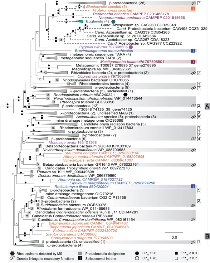
Figure 2.

Figure 2—figure supplement 1. Intron positions in eukaryotic rquA gene sequences.

Figure 2—figure supplement 1.

Nucleotide from both transcriptome and genomic sequencing projects (when available) were aligned using Sequencher (Gene Codes v 5.4.6) and introns were manually inspected for position, length and phase. The phase indicates the position of the intron relative to the codon where phase 0, 1 and 2 begin before the codon, in between position 1 and 2, or in between position 2 and 3 respectively. All introns were major spliceosomal introns with GT-AG boundaries. In general, intron positions and lengths were not conserved in distantly related species.
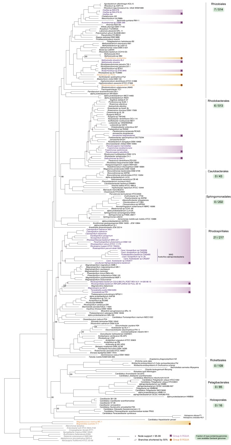
Figure 2—figure supplement 2. Phyletic distribution of RQUA among alphaproteobacteria superimposed on the alphaproteobacterial species tree.

Figure 2—figure supplement 2.

Maximum likelihood analysis of a 200 phylum-specific phylogenetic marker genes representing 54,400 sites from 210 representative alphaproteobacteria under the LG + C60 + F (PMSF)+ Γ4 model of evolution implemented in IQ-TREE. Tree was rooted using an outgroup of other proteobacteria. Nodes with maximal support are unlabeled, while those with support values between 95–99 are labeled with squares. RQUA-containing taxa are coloured in purple or orange representing Group A and Group B type RQUA respectively. Orders of alphaproteobacteria are indicated on the right of the tree with a summary of the number of alphaproteobacterial genomes interrogated for RQUA presence/absence in Genbank (green box).
Figure 2—figure supplement 3. RquA homologues lack critical S-adenosyl methionine (SAM) binding site.

Figure 2—figure supplement 3.

Conserved residues known to interact with the carbonyl group of SAM are shown with black triangles. A substitution from aspartate to glutamine was obversed in all bacterial (represented here by Rhodospirillum rubrum) and eukaryotic RquA demonstrated with a open circle. Taxa are coloured with super-Group Affiliation as in Figure 2. Motif features: [VILFG]-[LIVCS]-[DENLV]-[VALMIT]-[GLYCFA]-[CSTAYFPAG]-[GA]-[PTSGNKRMV]-[GD]).