
Figure 2. Male and female rats exhibit comparable primed hippocampal cytokine responses post-stress.
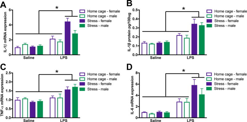
Male and female rats underwent inescapable stress or remained in the home cage. 24 h later rats received a single IP injection of 10 ug/kg LPS or saline and tissue was collected 3 h later. (A) Stress potentiated LPS-induced IL-1β mRNA elevations in the hippocampus of male and female rats. (B) IL-1β protein was similarly regulated in the hippocampus of male and female rats. Additional pro-inflammatory cytokine mRNA including (C) TNFα and (D) IL-6 were elevated by stress followed by LPS. Results were analyzed using 2 × 2 × 2 ANOVAs with sex, stress, and immune challenge as the between subjects factors (n = 6 – 8 per group with a total of 56 rats). Data are expressed as mean ± SEM. *simple effect of stress, p < 0.05 in all cases.