
Figure 3. Male and female rats exhibited comparable down-regulation of anti-inflammatory pathway genes following stress.
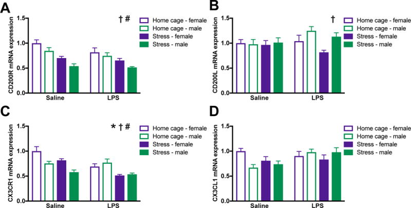
Male and female rats underwent inescapable stress or remained in the home cage. 24 h later rats received a single IP injection of 10 ug/kg LPS or saline and tissue was collected 3 h later. (A) CD200R mRNA was lower in the hippocampus of male as compared to female rats and was suppressed by stress in both sexes. (B) CD200L was increased in the hippocampus of male as compared to female rats and unaffected by and LPS stress. (C) CX3CR1 was reduced in the hippocampus of male as compared to female rats and suppressed by stress and LPS. (D) In contrast, mRNA expression of the CX3CL1 was unaffected by sex, stress, or LPS. Results were analyzed using 2 × 2 × 2 ANOVAs with sex, stress, and immune challenge as the between subjects factors (n = 6 – 8 per group with a total of 56 rats). Data are expressed as mean ± SEM. †main effect of sex, *main effect of LPS, #main effect of stress, p < 0.05 in all cases.