
Figure 4. Peripheral cytokine responses are potentiated in female but not male rats in response to stress and LPS.
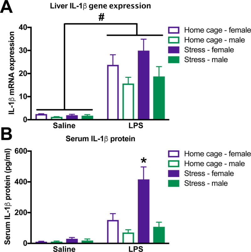
Female and male rats underwent inescapable stress or remained in the home cage. 24 h later rats received a single IP injection of 10 ug/kg LPS or saline and blood and tissue were collected 3 h later. (A) IL-1β mRNA expression in liver was increased by LPS but not stress in both female and male rats. (B) Serum IL-1β concentrations were increased by exposure to stress and LPS in female rats. In contrast, serum IL-1β was not affected by stress exposure in male rats. Results were analyzed using 2 × 2 × 2 ANOVAs with sex, stress, and immune challenge as the between subjects factors (n = 6 – 8 per group). Data are expressed as mean ± SEM. #main effect of LPS, *simple effect of stress, p < 0.05 in all cases.