
Figure 5. Microglia are primed by exposure to stress in male but not female rats.
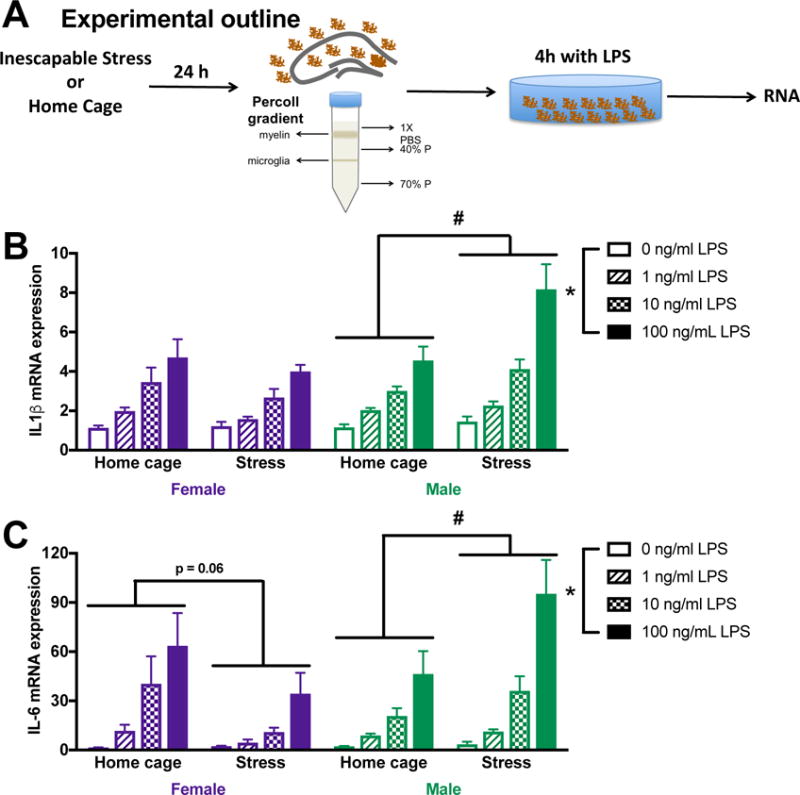
(A) Male and female rats underwent inescapable stress or remained in the home cage. 24 h later microglia were isolated from the hippocampus using a percoll density gradient. Cells were plated (10,000 cell per well) with LPS for 4 h prior to isolating mRNA. (B) Prior stress exposure potentiated IL-1β mRNA responses to LPS in microglia isolated from male rats. In contrast, IL-1β mRNA expression was unaffected by prior stress in female microglia. (C) Similarly, IL-6 mRNA expression was potentiated by stress exposure in males, which trends toward an overall suppression in microglia isolated from the female hippocampus. Results were analyzed using 2 × 2 × 4 ANOVAs with sex, stress, and LPS dose as the independent variables (n = 4 per group and experiment replicated in an additional cohort of 32 female rats [supplemental Fig 1]). Data are expressed as mean ± SEM. *main effect of LPS, #stress X LPS interaction, p < 0.05 in all cases.