
Figure 6. Stress reduces phagocytic capacity in microglia isolated from male and female rats.
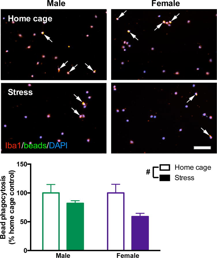
Male and female rats underwent inescapable stress or remained in the home cage. 24 h later microglia were isolated from the hippocampus using a percoll density gradient. Microglia were plated (5 × 10⁴ cells per well) with a latex bead solution (1:100 concentration). Microglia were identified with Iba1 (red), beads were conjugated to a green fluorophore, and nuclei were identified using DAPI (blue). Arrows highlight microglia that phagocytosed latex beads (yellow represents colocalization of red microglia and green beads). Stress caused fewer microglia to phagocytose the beads. Results were analyzed using a 2 × 2 ANOVA with sex and stress as the independent variables (n = 6 per group with a total of 24 rats; 20 images were analyzed per rat and averaged). Data are expressed as mean ± SEM. #main effect of stress, p < 0.05. Scale bar: 50 mm.