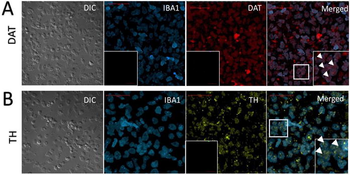
Fig. 2.

Monocytes constitutively express DAT and TH proteins. Confocal microscopy of immuno-stained monocytes isolated and cultured from human blood. A) DIC imaging (far left) of macrophages isolated and cultured from human blood are immuno-positive for IBA1 (second from left, with secondary Ab only labelled negative control inset), a macrophage-specific marker, and for DAT (third from left, with secondary Ab only labelled negative control inset). The same cells co-express both proteins (far right, inset); examples indicated by arrowheads in inset. B) DIC imaging of macrophages isolated and cultured from human blood are immuno-positive for IBA1 (second from left), a macrophage-specific marker, and TH (third from left, with secondary Ab only labelled negative control inset). Cells co-express both proteins, with zoomed-in examples indicated by arrowheads in far right inset. Scale bar is 50 μM; images are all representative of experiments repeated at least 5 times (n = 5).
