
Fig. 1. Characterization of prostate cancer cell-specific PBM nanoparticles for charge, size, morphology and surface chemistry.
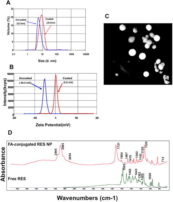
Resveratrol and docetaxel encapsulated; folic acid conjugated PCL-PEG coated and uncoated nanoparticles (A) size and (B) zeta potential (measured using a Malvern Zeta Sizer NS instrument). (C) Scanning electron microscope showing surface morphology of PBM nanoparticles coat with platinum–palladium layer. (D) Fourier transform infrared spectrum of synthesized resveratrol PBM nanoparticle and free resveratrol. The data were collected in absorption mode with 64 background scans and the wave number ranging from 4000 to 500 cm−1.