
Fig. 4. Z-stack orthogonal projections images to evaluate the targeted nanoparticle in prostate cancer cells.
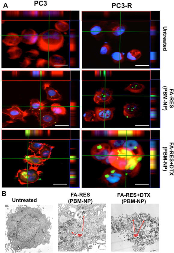
(A) Immunofluorescent staining of PCa cells treated with different dosages of green fluorescein labeled FA-conjugated RES-NP (3μM) and RES+DTX-NP (3μM+0.01μM) (Cells were immunostained with the anti-FRα antibody). Cells treated with NPs were detected by GFP and RFP (FRα), represented as green and red colors respectively. Nuclei were counterstained with DAPI (Blue). The orthogonal projections showed an abundant number of internalized combined nanoparticle (green) through folate receptor in PCa cells. Scale bar, 50 μm. (B) Internalization of PBM nanoparticles in docetaxel-resistant PC3-R cells treated with FA-conjugated RES-NP (3μM) and RES+DTX-NP (3μM+0.01μM), for 48 h and analyzed by TEM. The red arrow heads indicate the PBM nanoparticles within the cells.