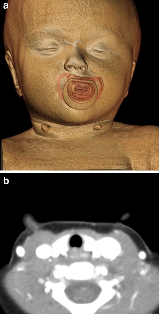
Fig. 2.

3D (a) and axial (b) CT images show tubular bilateral anterior lower neck lesions covered with skin and subcutaneous fat and a central intermediate attenuation cartilage core that extends to the underlying to the underlying sternocleidomastoid muscles
