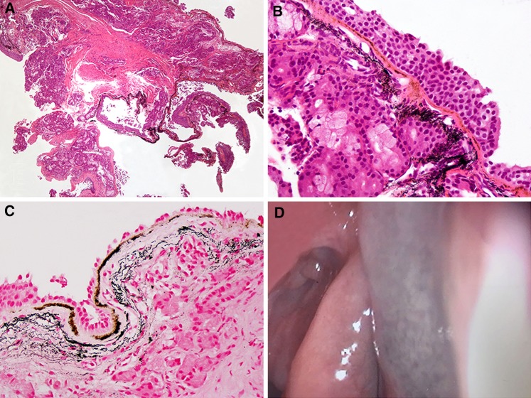
Fig. 1.
a Histopathological picture featuring an extensive subepithelial deposition of pigment. b The mucosal pigmentation characterized by a brownish subepithelial band associated with deeper diffuse cloud-like black granular deposits. Of note is the absence of any inflammatory cell infiltrate. c The same features as in (b), with Perls’ stain

