Fig. 1.
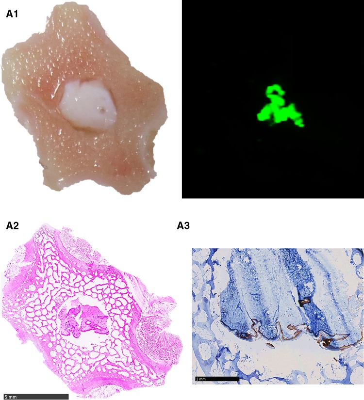
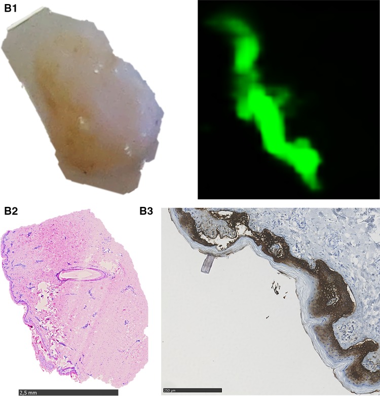
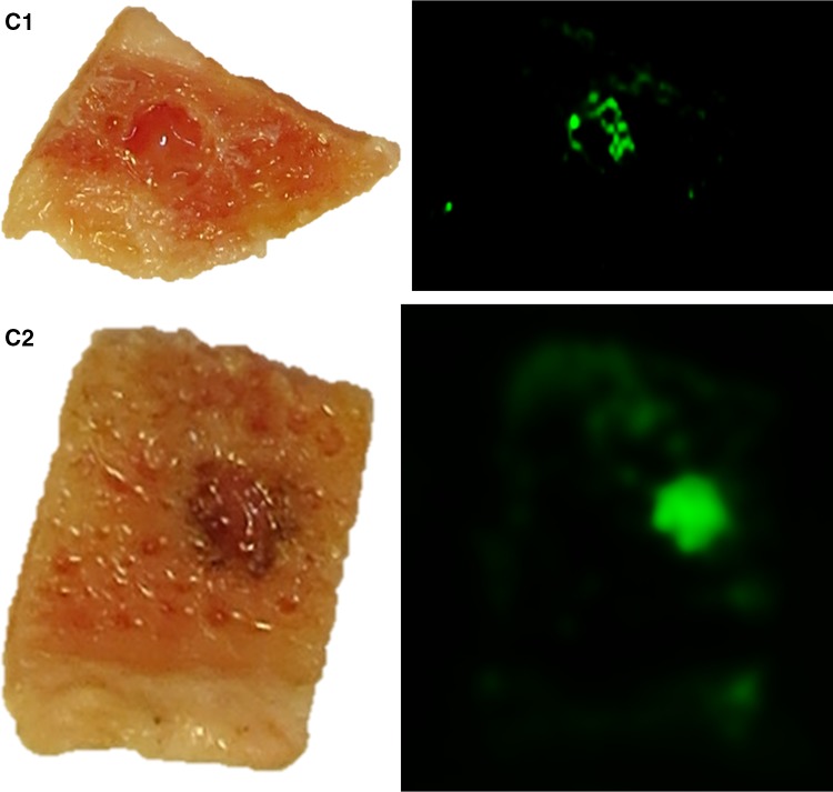
Representative results of the Tissue-ProtTrans method with composited bone wafers compared to the results of standard histological staining technique. A1 left part shows an image of native porcine bone wafer with skin placed on NC membrane immediately before protein transfer, right part shows the scanned membrane (800 nm laser beam) of this area after the Tissue-ProtTrans method; A2 H&E stain of the composite bone wafer after protein transfer, fixation, decalcification and paraffinisation; A3 consecutive IHC staining against CK 5/6 (5× fold magnified, selected part), B1 left part shows an image of porcine skin placed on NC membrane immediately before protein transfer, right part shows the scanned membrane (800 nm laser beam) of this area after Tissue-ProtTrans method, B2 H&E stain of the skin tissue after protein transfer, fixation, and paraffinisation. B3 consecutive IHC staining against CK 5/6 (5× fold magnified, selected part), C1 left part shows an image of human bone wafer with tumour tissue ø 4 mm placed on NC membrane right before protein transfer, right part shows the scanned membrane (800 nm laser beam) of this areal after the Tissue-ProtTrans method, C2 left part shows an image of human bone wafer with tumour tissue ø 2 mm placed on NC membrane right before protein transfer, right part shows the scanned membrane (800 nm laser beam) of this area after the Tissue-ProtTrans method


