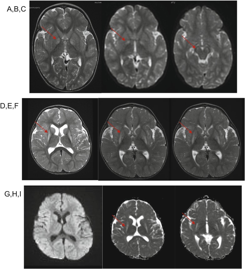
Fig. 2.
MRI for patient 1 (a–c) and patient 2 (d–i). Patient 1, images obtained at 2 years of age: (a) Axial T2-weighted MR demonstrates T2 hyperintensity of bilateral globus pallidi. (b, c) Diffusion-weighted imaging (DWI) demonstrates bilaterally restricted diffusion in globus pallidi and substantia nigra (arrows). Patient 2, images obtained at 14 months of age: (d–f) Axial T2-weighted MRI demonstrates T2 hyperintensity of bilateral putamina, and to a lesser degree globus pallidi (arrows). (g) Diffusion-weighted imaging (DWI) and (h, i) apparent diffusion coefficient (ADC) sequences demonstrate that regions of signal change in putamina and globus pallidi lack restricted diffusion

