Figure 2.
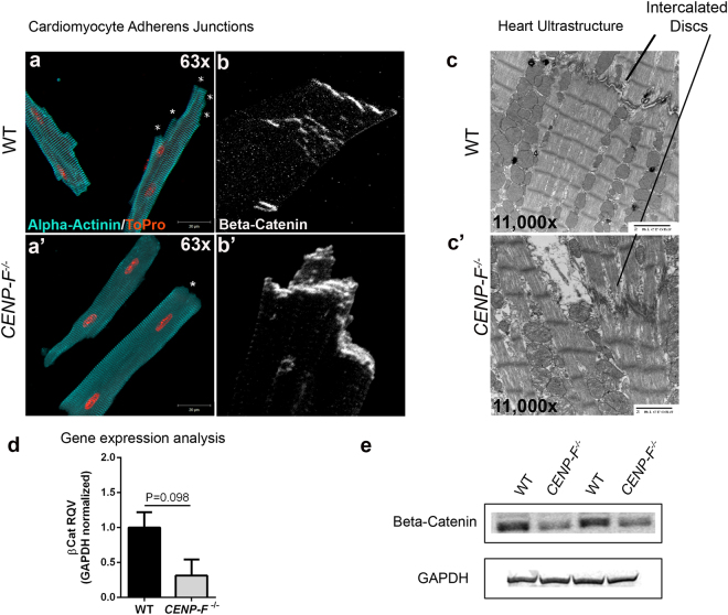
Intercalated disc organization is disrupted with the loss of CENP-F in cardiomyocytes. Cardiomyocytes isolated from wild-type (a) and CENP-F−/− (a’) mice were fixed and immunostained with alpha-actinin 2 antibody (teal) and TOPRO (orange). In CENP-F+/+ cardiomyocytes, there are multiple junction ends, as indicated by * (a). However, in CENP-F−/− cardiomyocytes, the cell termini is blunted, indicated by * (a’). The intercalated discs were immunostained with beta-catenin antibody (white) and the images are zoomed in to depict the lining of the cardiomyocyte junctions. In wild-type mice, there is thin and distinct junction staining (b) opposed to disintegrated and thickened junction staining of the mutant cardiomyocytes (b’). An EM image of wild-type heart shows a clear depiction of an intercalated disc, see horizontal structure pointed to by line (c), In CENP-F−/− cardiomyocytes, the intercalated disc is highly disintegrated (c’). A gene expression analysis of beta-catenin in wild-type versus CENP-F−/− cardiomyocytes (d). A western blot analysis of beta-catenin in wild-type versus CENP-F−/− cardiomyocytes showed significant decrease in beta-catenin. 2 cropped samples are presented and n = 5, p < 0.05. (Full-length gels are presented in Supplementary Fig. 4). Scale bars: (a) 20 mm; (a’) 20 mm; (c) 2 microns; (c’) 2 microns. Error bars in (e) represent standard error of the mean (SEM).

