Figure 3.
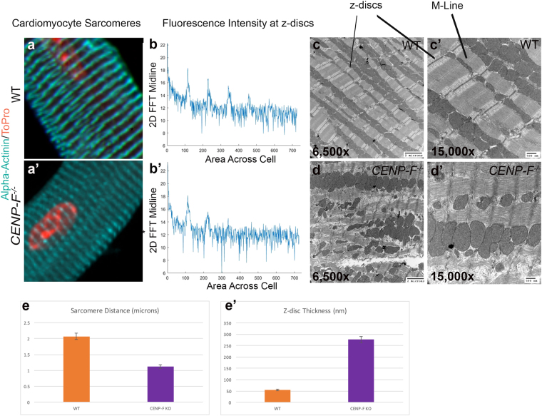
Sarcomere architecture is disrupted with the loss of CENP-F in cardiomyocytes. Cardiomyocytes isolated from wild-type (a) and CENP-F−/− (a’) mice were fixed and immunostained with alpha-actinin antibody (teal) and TOPRO (orange). A zoom in view of the sarcomere structure displayed a distinct patterning in wild-type cardiomyocytes (a) versus a diffuse and thickened z-disc patterning in CENP-F−/− cardiomyocytes (a’). A 2-D spectrum analysis of the fluorescence regularity in alpha-actinin stained cardiomyocytes displayed high intensity points at regular positioning in wild type cardiomyocytes (b). There is much more variability in the fluorescence staining of the CENP-F−/− cardiomyocytes (b’). A TEM of a wild-type adult mouse heart shows the overall precise patterning of sarcomeric structure (c). The z-discs and M-lines are well defined, as depicted in a higher magnification (c’). In a CENP-F deleted mouse heart, the z-discs are disintegrated and there are breaks within the actomyosin network (d,d’). The distance between the z-discs is much shorter in CENP-F−/− cardiomyocytes in comparison to the wild-type population, *p < 0.001 (e). Additionally, the width of the z-discs is much greater in the mutant cardiomyocytes opposed to the wild-type, *p < 0.001 (f).Scale Bars: (c) 2 microns; (c’) 500 nm; (d) 2 microns; (d’) 500 nm. Error bars in (c,d) represent standard error of the mean (SEM).

