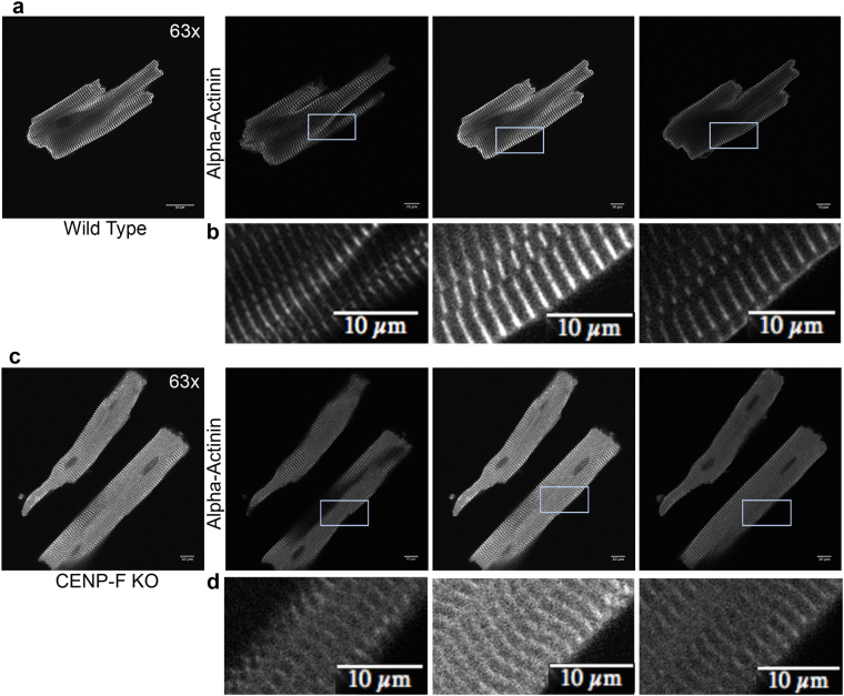
Figure 4.
CENP-F−/− cardiomyocytes have a disorganized z-disc structure compared to wild type cardiomyocytes. Individual slices of z-stack images in wild type cardiomyoctes show distinct z-disc staining with anti-alpha-actinin antibody (a,b). In CENP-F−/− cardiomyocytes, alpha-actinin staining is not sharp/distinct. Additionally, z-discs are widened and severely disintegrated (c,d). This is in agreement with quantification of z-disc width conducted on TEM images (see Supplementary Fig. 4) All images are adjusted at the same brightness and contrast levels. Images are in greyscale for proper comparison of image quality. Scale bars: (a) 20 um; (b) 10 um; (c) 20 um; (d) 10 um.

